




Dr. Heather Duncan
DVM DAVDC
Crown Veterinary Dental Specialists
Courses by Dr. Heather
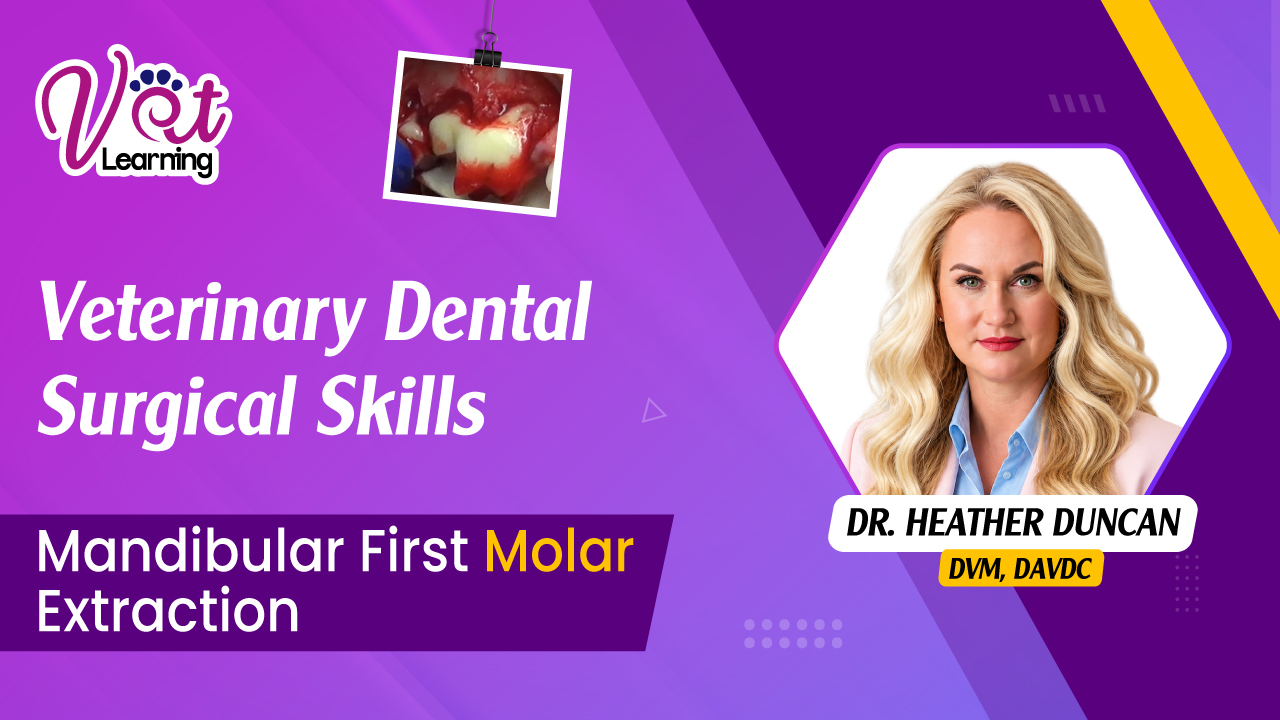
Mandibular First Molar Extraction
A practical, step-by-step guide to extracting the mandibular first molar in dogs, from flap creation and tooth sectioning to extraction and smooth, tension-free closure.
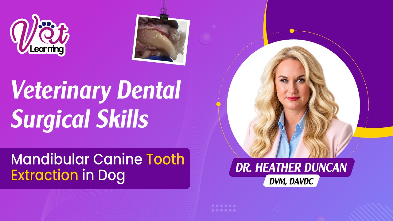
Mandibular Canine Tooth Extraction in Dog
This course provides a practical look at mandibular canine extraction using a canine specimen. It walks through flap creation, removal of alveolar bone, tooth elevation, and management of the surgical site, with a focus on protecting surrounding tissues and achieving a smooth, tension-free closure to support proper healing.
Veterinary Dental Surgical Skills
A systematic, step-by-step approach to exposure, controlled luxation, and tension-free closure in the dog.
Canine - Maxillary Second Molar Extraction & Flap Closure
Course Summary This comprehensive clinical training program provides an in-depth, step-by-step masterclass on the surgical extraction of the canine maxillary second molar and the subsequent execution of tension-free mucoperiosteal flap closures. Due to the unique, delicate anatomical location of the maxillary second molar—positioned directly ventral to the orbit and retrobulbar space—surgical intervention in this area carries heightened risk for severe complications, including root displacement, orbital trauma, and flap dehiscence. This course bridges fundamental oral surgery theory with practical, real-world execution, guiding clinicians through evidence-based protocols that prioritize tissue preservation, structural safety, and long-term surgical success. Through a detailed four-module structure combining high-definition surgical video breakdowns, anatomical risk mapping, step-by-step procedure guides, and rigorous clinical assessments, practitioners will develop the technical competence and confidence required to safely extract multi-rooted upper molars, perform tooth sectioning, release periosteal tension, and execute advanced suturing techniques. Core Learning Objectives Upon successful completion of this course, clinicians will be able to: Perform Anatomical Risk Audits: Identify key anatomical landmarks surrounding the canine maxillary second molar, specifically the orbit, retrobulbar space, major palatine artery, and infraorbital neurovascular bundle, to prevent intraoperative complications. Execute Safe Elevation Dynamics: Implement controlled rotational and luxating leverage using a 3C elevator with a mandatory short finger stop to minimize slippage and eliminate accidental orbital penetration. Master Surgical Tooth Sectioning: Utilize high-speed burs under continuous irrigation to section multi-rooted molars along natural furcation lines when simple elevation yields minimal tooth mobility. Identify Apical Pathology: Recognize endodontic and periodontal pathology, such as apical granulomas, root resorption, or fractured root tips, upon removal of root segments. Design and Mobilize Mucoperiosteal Flaps: Design full-thickness pedicle flaps and execute precise periosteal releasing incisions to eliminate surgical line tension. Apply Advanced Oral Closure Patterns: Select appropriate absorbable suture materials (e.g., 4-0 Monocryl) and perform simple interrupted and circumferential (purse-string) suturing techniques around adjacent anchor teeth to ensure complete edge apposition and prevent post-operative dehiscence. Modules Module 1: Initial Assessment and Elevation Focuses on mobility assessment, luxator placement, leverage principles off the maxillary first molar, and safety mechanics for protecting the adjacent eye. Module 2: Tooth Sectioning and Root Extraction Demonstrates the step-by-step separation of the palatal cusp using a #2 round bur under water cooling, safe fragment extraction, and avoiding deep, unguided root retrieval in the maxillary alveolus. Module 3: Mucosal Flap Design and Release Covers the design of surgical flaps, elevation of mucoperiosteum from caudal bone, and periosteal release incisions using LaGrange scissors to achieve complete, tension-free mobility. Module 4: Flap Closure and Suturing Concludes with surgical closure techniques, detailing knot security, 1–2 mm tag trimming, simple interrupted patterns, and specialized circumferential suturing around M1.


Free Webinars (upcoming)
Past Webinars
About Me
About Heather Duncan
Growing up in North Carolina, at a young age, Dr. Heather Duncan decided that she wanted to become a veterinarian to help her pets that she loved so much as well as other people’s furry family members. She graduated Magna Cum Laude from North Carolina State University and then received her DVM from Purdue University’s School of Veterinary Medicine in 2005.
After graduating veterinary school, Dr. Duncan practiced at a progressive 24-hour small animal hospital in Saint Petersburg, FL. Realizing her passion for Veterinary Dentistry, she began a three-year Veterinary Dentistry and Oral Surgery Residency program in 2008 at the Animal Dental Center of Baltimore and Annapolis, MD. Dr. Duncan developed the Dentistry & Oral Surgery Service at Upstate Vet Emergency & Specialty Care in Greenville, SC in 2013, then went on to become a Diplomate of the American Veterinary Dental College (AVDC) and the first Board Certified Dentist in South Carolina in 2014.
She enjoys all aspects of veterinary dentistry (Endodontics, Fracture repairs, Oral Surgery, Orthodontics, Periodontics and Restorative dentistry).
Dr. Duncan is active in the American Veterinary Dental College, volunteering on many committees. She thoroughly enjoys teaching throughout the United States and is passionate about sharing her knowledge of Veterinary Dentistry.